


Warts are formations on the skin in the form of nodules or papillae. It is the most common skin pathology, occurring in more than 90% of the world's population. Warts can appear on any person, at any age, on absolutely any area of the skin, from the face to the feet. The disease is often contagious, everything depends on the person's immune system.
What causes warts
There is a common belief that touching a frog causes warts to appear. This is a fallacy. The causative agent of the disease, which leads to the formation of warts, is a human papillomavirus infection. According to statistics, this infection causes about 20% of all cancers.
The risk of HPV infection increases significantly:
- when using items for personal hygiene and items for general use of other people;
- in public places (swimming pool, bathroom, etc. ), especially when you go there barefoot;
- in case of skin damage;
- with increased sweating of hands and feet;
- in contact with an infected person (handshake, sexual contact, etc. );
- when walking in tight, uncomfortable shoes that cause friction on the skin of the feet;
- when using non-sterile tools (in a beauty salon, etc. ).
Are warts always dangerous?
Most warts are completely harmless and can theoretically disappear within a few weeks or at most a month. In this case, patients are more likely to worry about a serious cosmetic defect that causes psychological discomfort and prevents them from leading a fulfilling lifestyle.
Warts are often painless unless they are on the soles of the feet or another part of the body that is subject to impact or constant contact. But there are cases of itching and discomfort in the affected area.
How to recognize warts: symptoms and signs
An inexperienced person can confuse warts with other skin formations, for example, moles, calluses, melanomas.
The main differences between warts and moles:
- moles have a dark or black shade, while warts have a light color;
- warts grow tightly together with the skin, moles are separate structures, as if glued to the body;
- moles are soft and smooth to the touch, warts are hard, firm and rough.
In addition, it is easy to distinguish a wart from a callus. When pressure is applied to the growth, painful sensations appear, and if it peels off, traces of bleeding will be visible under it. Beneath the callus is new, tender skin.
You can tell a wart from a melanoma by color and shape. This dangerous disease is characterized by heterogeneous red and black shades, proliferation and uneven contour.
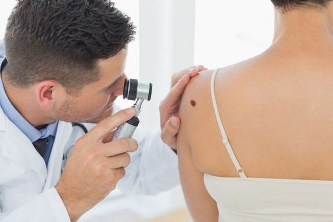
It is not difficult for a dermatologist to make the correct diagnosis using a visual examination. But a good specialist will not be satisfied with just a simple examination. He will certainly use a special magnifying device - a dermatoscope. If a pathogenic process is suspected, scraping of the surface layer will be necessary.
Anogenital warts (located around the anus and on the genitals) require consultation with a gynecologist or proctologist.
What is the structure of benign neoplasms
Growths consist of cells that have partially retained their original functions and are capable of slow growth. They are similar in structure to the tissues from which they are derived. They can exert pressure on nearby tissues, but do not penetrate them, since they have a capsule in their structure. They respond well to hardware and surgical treatment and, as a rule, do not cause relapses.
There are always congenital formations on the skin - moles or warts, as well as acquired ones. The latter are formed on the surface or in the subcutaneous tissue as a result of metabolic disorders, reduced immunity or under the influence of a virus.
Common (simple, vulgar) warts
Common warts are thick, dry growths characterized by an uneven and rough surface to the touch, variable size and rounded shape. They appear as a hard, keratinized balloon up to 1 cm in diameter, significantly raised above the surface of the skin.
The surface of ordinary warts is often covered with depressions and protrusions, which is why the neoplasm vaguely resembles a cauliflower or raspberry with black dots inside.
This is the most common type of wart, accounting for up to 70% of all such skin neoplasms. Common warts can appear on the skin at any age, but most often affect children and young adults. This is due to the fact that they have weaker immunity than adults.
Common warts usually appear on the hands (fingers and backs of the hands), knees and elbows, sometimes on the face or legs, and extremely rarely on the lining of the mouth.
Scattered small growths can form next to the large "parental" wart. Young neoplasms usually remain flesh-colored, over time they acquire a dirty gray or gray-brown hue, less often yellow or pink. This is due to their uneven porous surface, which accumulates dirt.
Vulgar warts usually do not cause concern: they do not cause unpleasant symptoms, do not hurt and do not itch. However, they can cause pain if they are in areas subject to impact or in contact with clothing. Growths can heal on their own over time, especially if they appear in childhood.
Why do benign growths appear on the skin?
Cosmetologists and dermatologists do not know the exact mechanism of their formation. Most often the reason is:
- injuries;
- viruses;
- systemic diseases of the body, for example, xanthomas, arise due to an excess of fat in the blood;
- long-term skin diseases;
- exposure to aggressive substances;
- excessive exposure to ultraviolet radiation;
- X-ray;
- heredity (for example, seborrheic dermatosis).
Most skin lesions are benign
Plantar (spike) warts
Plantar warts are a type of vulgar wart. The manifestation of the disease is observed most often in children and at the age of 20-30 years. Of all skin warts, plantar warts occur in 30%.
Plantar warts appear as hard, round lumps with papillae in the middle. Characteristic black dots are visible inside the wart - numerous small thrombosed capillaries. At the edges there is a small roll of keratinized skin. The visible part, rising above the surface of the skin by only 1-2 mm, can reach 2 cm in diameter and is only a quarter of the total size of the plantar wart, which is formed mainly in the deep layers of the epithelium (skin).
Externally, the spine resembles a callus. A plantar wart can be differentiated (distinguished) from a callus by a visible break in the skin pattern consistent with the wart.
This type of neoplasm usually affects the feet (foot, sides and toes) and less often the palms. They appear on the skin as small whitish, point-like skin lesions, sometimes itchy. Over time, their surface becomes rougher and changes color - from yellow to dark brown.
Plantar warts themselves do not pose a threat to health, but when walking they cause a person significant discomfort, cause pain that often increases and may even bleed. This is due to the location of the tumor and the specificity of its growth. As the spine grows inward, the weight of the body when walking compresses the pain receptors.
The incubation period of the disease varies from several days to several years. The infection enters the body and goes into standby mode to activate a favorable environment. Plantar warts regress without treatment in 50% of cases. But this process lasts from 8 months to a year and a half.
Without treatment, plantar warts will enlarge and multiply, even to the point of forming large clusters of tumors. It can even cause a person to be temporarily incapacitated due to excruciating pain that prevents them from walking.
Based on the characteristics of the lesion and its location, plantar warts are divided into 3 types:
- just;
- periungual;
- mosaic.
Do benign growths hide the danger?
Benign neoplasms are unpredictable structures that can appear at any time or not at all. The process of their transformation into malignant is not fully understood. There is no unequivocal answer to the question of what exactly activates this process. Mechanical trauma, excess ultraviolet radiation, metabolic disorders, and other factors are thought to contribute to degeneration. One way or another, if you have a benign skin lesion, you should not experiment and rely on chance. In addition, today the removal does not cause difficulties.
Periungual plantar warts
Periungual warts are small, rough formations with cracks on the surface, located on the hands and feet of a person, namely near the nail plate or deep under it. Outwardly, they look like heads of cauliflower.
They can be flat, pointed or hemispherical. As a rule, perianal warts are gray, but they can also be flesh-colored. They are not too dense, like ordinary plantar, but have a rather deep root.
This disease mainly affects children and young people. The main factor for infection are skin microtraumas around the nail. At particular risk are those who bite their nails and pet stray animals, as well as people who carelessly remove cuticles, use unsanitized tools, and work in water without gloves.
This type of neoplasm does not pose a threat to human health, it is mostly just a cosmetic defect. Periungual plantar warts do not cause discomfort or pain on pressure. However, the wart under the nail is not so harmless - over time, the neoplasm provokes exhaustion of the nail plate and its further destruction.
In addition, various bacteria and viruses enter through cracks on the surface of growths, which are easily formed due to frequent hand work, causing re-infection. Also, when the warts grow, the cracks can cause pain. The cuticle is often lost and a tendency to inflammation (paronychia) develops.
Removal of the tumor is necessary to stop the proliferation of growths that easily spread to healthy fingers. The localization of the wart under the nail plate significantly complicates treatment and removal. When it appears in childhood or adolescence, it may disappear on its own.
Where do warts come from - they are contagious!
Like herpes, warts are the result of a virus. More than a hundred types of viruses are responsible for the development of warts, most of which are HPV. Since there are oncogenic types of HPV, some formations can be particularly dangerous in terms of cancer, for example, those that develop around the genitals.
No matter what the warts are or where they are, never scratch, rub or scratch them as they can transmit millions of viruses to other areas of the skin where new growths can appear!
It is very easy to get infected with wart viruses. For example, infected human epithelial cells fall into the pool water. They swim in the water and easily find their prey. The wart virus can also be spread through direct physical contact, simply by shaking hands. The penetration of viruses into the body is facilitated by small lesions on the skin.
In children, warts often appear under the nails as a result of finger sucking or chewing, which can be painful and difficult to treat. Children can easily pick up viruses while playing. As a result, every fourth child has viral warts on their hands or feet.
Whether we get the virus depends on how strong our immune system is. A strong immune system suppresses the infection that causes warts.
Mosaic plantar warts
Mosaic warts are a special type of neoplasm. They are plaques, so-called clusters, formed as a result of the fusion of many small plantar warts tightly pressed together. The arrangement of the plates resembles a mosaic (hence their name).
This formation is usually observed in a small and localized area. It can reach a diameter of about 6-7 cm. In the early stages of development, mosaic warts look like small black holes. As they develop, they take on the appearance of a white, yellowish or light brown cauliflower, with dark spots in the middle. These spots are formed due to thrombosis of blood vessels.
This type of wart is quite rare. They usually affect the hands or soles of the feet and are especially common under the toes. Unlike common plantar warts, mosaic warts cause little or no pain when walking because they are flatter and more superficial.
Mosaic warts are highly contagious. They are difficult to treat because of the multiple foci of viral infection. The success of treatment is facilitated by its timely initiation. As a rule, mosaic growths are prone to recurrence even after surgical removal.
Benign and malignant skin neoplasms: what are the differences?
Benign pathologies do not pose a threat to human life. If they reach large sizes, they can interfere with the proper functioning of various body systems. In contrast, malignant ones grow quickly and aggressively, penetrate the surrounding tissues and form metastases over time. Some damage vital organs and cause death.
Sometimes benign skin tumors change due to external or hereditary reasons. They acquire the ability to degenerate into malignant pathologies. Such conditions are called borderline or precancerous. They pose a great danger to health and life, although they do not always have pronounced symptoms.
Flat (juvenile) warts
Flat warts are a fairly common type of tumor and the least problematic. They present as small lenticular lesions (a few mm in diameter) or smooth papular lesions. They can grow either individually, which is quite rare, or in large numbers, close to each other.
There are several stages of the disease:
- mild - one or several painless warts;
- medium - from 10 to 100 painless formations;
- severe - more than 100 neoplasms.
If they are located in places that experience excessive pressure (friction from clothes, shoes, etc. ), they cause pain.
Flat warts are easily recognized and have a white, brown, yellowish or pinkish tint similar to the color of the flesh. They are about the size of a pinhead and compared to other types of warts, they are smoother and flatter. In fact, where a flat wart develops, the skin is slightly raised (at a height of about 5 mm), forming a kind of raised circular area.
The growths usually appear on the face, knees, elbows, back, legs and hands (especially the fingers). People of absolutely any age become victims of this disease. But it most often affects children and adolescents (20% of schoolchildren have it), hence the second name of warts - juvenile.
In a close group of students, 80% showed resistance (resistance) to the virus. In adults, irritation and inflammation after shaving contribute to the proliferation of tumors.
The incubation period of the infection can last up to 8 months. Most often, the disease is only a cosmetic defect. Juvenile warts are painless unless caused by mechanical pressure or injury and can sometimes itch, but are extremely contagious.
The virus is practically not transmitted through common objects, the main route of infection is skin contact. Flat warts multiply so easily that it is enough to touch a healthy part of the body to cause the birth of a new formation.
The peculiarity of this type of warts is that in most cases no treatment is required: they can disappear as suddenly as they appeared, especially in children. In adults, the disease must be treated, and the virus is very resistant to drug treatment.
Transmission of warts by direct contact
Mild trauma or maceration leads to epithelial barrier dysfunction and subsequent loss of skin integrity, opening the way for viral infection and wart formation. The incubation period ranges from 3 weeks to 8 months after exposure. Spontaneous regression is observed in most cases.
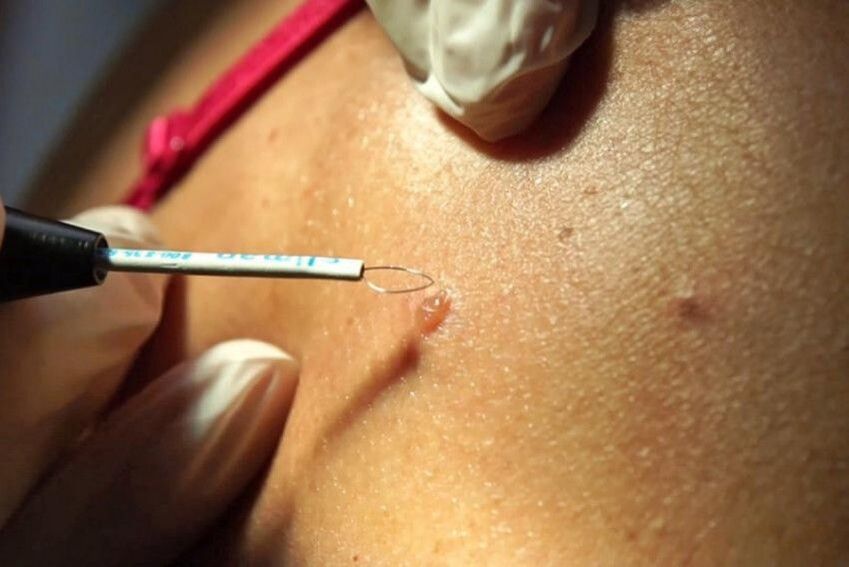
Laser wart removal
Today, laser surgery is one of the best ways to get rid of warts. It is a painless and safe procedure that can be used in areas of maximum sensitivity. Laser removal of tumors is very effective: the probability of recurrence is minimal. This is significantly affected by the severity of the disease.
Warts are removed by layer-by-layer burning of the affected area, thanks to which the doctor controls the depth of the effect. At the same time, the laser beam burns the blood vessels, thereby preventing bleeding at the site of exposure.
Three methods of laser coagulation are common:
- Carbon dioxide (CO2) laser. Procedures with this laser are more painful. Although the CO2 laser seals the blood vessels, it also kills the wart tissue. In this process, there is a possibility of damage to healthy tissue. The wound usually takes longer to heal and scarring is possible. The efficiency is about 70%.
- Erbium laser. It is characterized by a shorter wavelength. The likelihood of scarring after healing is greatly reduced.
- Pulsed dye laser. This laser more effectively seals the blood vessels that feed the wart. It does not damage much of the healthy tissue like the CO2 laser does. It is also the only type of laser approved for use in children. The effectiveness of this method of treatment is about 95%.
| Advantages | disadvantages |
| Minimal probability of scar formation (depending on the degree of neglect of the pathology) | High price |
| Fast tissue healing | |
| High efficiency of the method | |
| Minimal damage to healthy tissue | |
| Speed of the procedure |
Wart removal is performed under local anesthesia. A crust remains at the cauterization site, which disappears within 14 days. After the procedure, the patient quickly returns to his normal lifestyle, subject to all the doctor's recommendations.
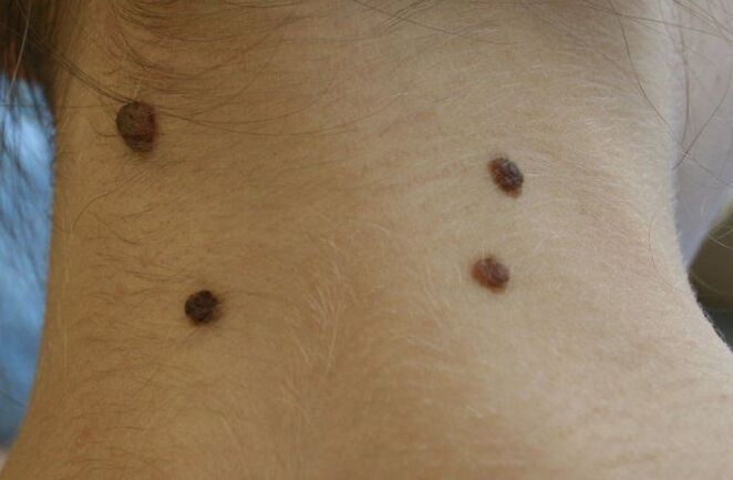
Treatment of filiform papillomas
In 90% of cases, filiform warts do not heal on their own (just as juvenile or vulgar warts in childhood can heal on their own).
They need to be treated. Especially if these formations are injured.
For example, if the papilloma is on the neck, it can be injured by a chain or collar of clothes. If on the face - from glasses, under the chest - from a bra. You should know that such permanent damage can lead to inflammation of this entity and its pain.
Official methods and methods of treatment
Laser removal of filiform warts - read a detailed article on laser removal.
The simplest and fastest, but cheap way to treat this type of papillomas. The doctor directs the laser beam at the skin formation, which vaporizes and burns it. First you need to anesthetize the skin with novocaine so that the patient does not feel pain. And wear safety glasses on your eyes.
The whole procedure takes no more than 1 minute per wart. The consequences are a small crust on the wound. After 3-5 days, this crust comes off and healthy and clean skin is formed in this place.
Removal by radio wave method - read the article about radio wave surgery.
The principle of operation is as follows: a device for radio wave surgery ("Surgitron") creates a high-frequency radio wave that destroys the wart tissue in the same way as a laser, i. e. vaporizes it.
The entire procedure is carried out in the same sequence as in the laser treatment method - first (mandatory! ) local anesthesia, then exposure for 1 - 2 minutes (it all depends on the size of the tumor to be removed). The consequences of the radio wave treatment are exactly the same as those of the laser.
Removal of filiform papillomas with liquid nitrogen - read information about liquid nitrogen.
This method is popular because of its simplicity. No need to numb the skin with injections, no need for a doctor to be present. The procedure can be performed by any nurse or employee in a cosmetic clinic.
Principle of action: liquid nitrogen with a temperature of minus 195 degrees freezes the tissue of the wart. A doctor or nurse, by dosing the effect on the skin over time, does not allow freezing of adjacent healthy areas of the skin around the pathological formation.
After completion of the procedure, in 90% of cases, papillomas disappear on their own within 3-4 days.
Electrocoagulation of filiform warts.
Nowadays, this method is used much less frequently, as it is a more traumatic method. Papillomas are cut out with an electric knife. In this case, a burn and a wound are formed on the skin, which then take longer to heal.
Radioknife removal
The most effective modern method of removing warts is radio wave removal. First of all, this is due to the fact that in this procedure the instruments do not come into contact with the patient's body: they are produced at a radio frequency.
Other advantages of radio wave wart removal should be noted:
- complete painlessness;
- speed of the procedure;
- exclusion of edema and infiltration;
- lack of postoperative complications;
- absence of scars at the site of wart removal;
- quick rehabilitation period.
The procedure is also performed under local anesthesia. After exposure, a crust forms on the affected area of the skin, which disappears on its own within 7-10 days.
Prevention of skin tumors
Unfortunately, medicine has not yet learned to prevent the appearance of various formations on the skin. But dermatologists give their patients the following preventive recommendations:
- do not delay in contacting a doctor if a tumor appears on the skin;
- removal of formations only after a specialist and diagnostics confirm their benign nature;
- avoid excessive exposure to the open sun;
- use sunscreen products, especially if you are prone to moles and hyperpigmentation;
- do not come into contact with chemically active and carcinogenic substances;
- do not eat foods that contribute to the development of cancer (smoked meats, sausages, animal fats, meat products with food stabilizers).